Advanced Treatment for Cholecystitis and Gallbladder Adhesions
The Gallbladder Is It Truly "Good"? (A Case Study on Cholecystitis)
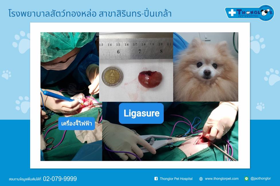
 Nong Cake was brought to the hospital presenting with lethargy and anorexia (loss of appetite). Following a physical examination and blood work, she was referred for an abdominal ultrasound to evaluate her internal organs.
The diagnostic imaging revealed Cholecystitis (inflammation of the gallbladder). The gallbladder was distended and filled with significant biliary sludge and sediment. Typically, a healthy gallbladder should not exceed 4 cm in size, with a wall thickness of less than 3 mm. In Cake’s case, the gallbladder had started to develop fibrous adhesions, attaching itself to the liver—a critical condition that increases the risk of gallbladder rupture if left untreated.
Surgical Intervention and Advanced Technology
To ensure the highest level of safety, a surgical removal of the gallbladder (Cholecystectomy) was performed. Due to the adhesions to the liver, our surgical team utilized specialized equipment:
- Electrosurgery: Used to minimize blood loss (hemostasis) and prevent infection by sealing tissues and blood vessels simultaneously as they are cut.
- LigaSure™: A state-of-the-art vessel-sealing technology that uses a combination of pressure and electrical energy to permanently fuse and cut small to medium-sized blood vessels with extreme precision.
The Medical Team:
- Attending Veterinarian & Anesthesiologist: Dr. Ukritkarn Konglertmongkol
- Radiologist (Ultrasound): Dr. Pornpimon Chanwanitchatrakul
- Surgeon: Dr. Peeraya Punnuwong
"For any inquiries regarding pet care, grooming, swimming sessions, or online shopping, feel free to reach out to us through the following channels:
- Tel: 02-079-9999
- LINE Official: @jaothonglor or click LINE @jaothonglor
- Facebook: Thonglor Pet Hospital
- Book an Appointment: tlpet.club/Thonglor-appointment
- Shop Online: tlpet.club/petshop"
#ThonglorPetHospital #TheBestAlways
Related in This Category
The Deadly Crunch Why Chicken Bones are a Life-Threatening Threat to Your Dog
Essential or Optional? Understanding Vaccines, Parasite Prevention, and Deworming for Dogs

When a Tiny Lump is No Small Matter
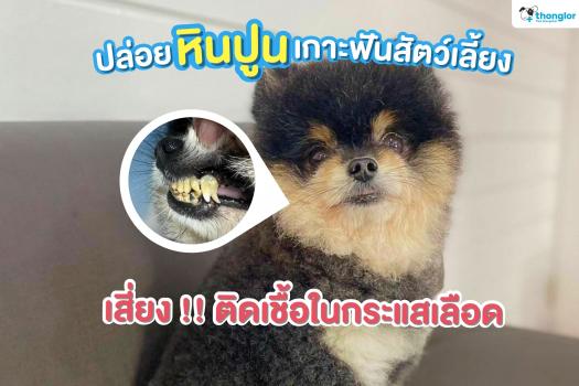
A Hidden Path to Bloodstream Infection