Cloudy Eyes in Dogs: Cataracts or Normal Aging?

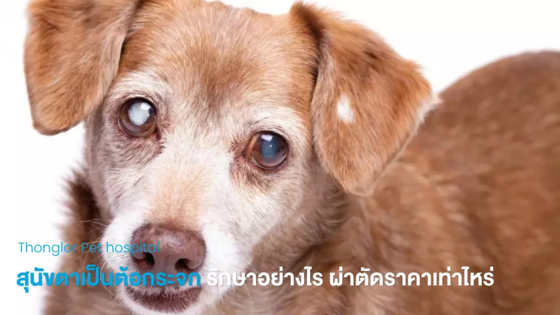
Cloudy Eyes in Dogs: Cataracts or Normal Aging? Symptoms, Treatment and Surgery
When an older dog’s eyes begin to look cloudy, gray or white, many owners wonder whether this is simply a normal sign of aging or whether their dog has developed cataracts.
Cloudy eyes can have several causes, including normal age-related changes of the lens, cataracts, corneal disease, inflammation inside the eye and glaucoma. Because these conditions require different approaches to treatment, an eye examination is important for determining the cause.
If your dog develops cloudy eyes together with bumping into objects, hesitation when using stairs, difficulty locating food or toys, eye redness, squinting or signs of eye pain, an ophthalmic examination should be considered.
Are Cloudy Eyes in an Older Dog Cataracts or Normal Aging?
One condition commonly mistaken for cataracts is nuclear sclerosis, also called lenticular sclerosis.
Nuclear sclerosis is an age-related change in the lens that commonly occurs in older dogs.
| Feature | Cataracts | Nuclear Sclerosis |
|---|---|---|
| Appearance | White or opaque areas within the pupil, affecting part or all of the lens | Usually a gray or bluish haze inside the eye |
| Vision | Vision may decrease depending on the location and severity | Most dogs can continue to see and function normally |
| Treatment | Surgery may be considered when vision is affected | Surgery is generally not required |
| Diagnosis | Requires an ophthalmic examination | Must be differentiated from cataracts and other eye diseases |
Cataracts and nuclear sclerosis can look similar from the outside. A photograph or visual inspection alone may not be enough to distinguish between them accurately.
What Causes Cloudy Eyes in Dogs?
Cloudy eyes do not always mean cataracts.
Possible causes include:
Cataracts – loss of transparency of the lens, reducing the amount of light that reaches the retina
Nuclear sclerosis – age-related changes within the lens
Glaucoma – increased pressure inside the eye that may cause cloudiness, enlargement and pain
Uveitis – inflammation inside the eye that may cause redness, discomfort and secondary lens changes
Corneal disease – cloudiness affecting the clear surface at the front of the eye rather than the lens itself
Because each condition is treated differently, identifying the location and cause of the cloudiness is important.
What Are the Signs of Cataracts in Dogs?
Signs that may be associated with cataracts include:
White, gray or cloudy appearance within the pupil
Bumping into furniture or other objects
Hesitation when going up or down stairs
Reduced confidence in unfamiliar environments
Difficulty locating food or toys
Becoming startled when approached from the side
Rapid development of cloudy eyes in dogs with diabetes
If your dog has redness, squinting, obvious eye pain, an enlarged eye or suddenly reduced vision, the eye should be evaluated promptly because inflammation or glaucoma may also be present.
What Are the Four Stages of Cataracts in Dogs?
Cataracts are commonly classified according to how much of the lens is affected.
| Stage | General Appearance | Effect on Vision |
|---|---|---|
| Incipient | Small or localized areas of lens opacity | Vision may be minimally affected |
| Immature | A larger portion of the lens becomes cloudy | Vision may begin to decrease |
| Mature | Most or all of the lens becomes opaque | Vision is usually significantly reduced |
| Hypermature | The cataractous lens begins to undergo further degeneration | Risk of inflammation and other complications increases |
The stage of a cataract is only one part of treatment planning.
Veterinarians will also evaluate retinal function, eye pressure, inflammation, other eye abnormalities and the pet’s overall health before determining whether surgery is appropriate.
Can Cataracts in Dogs Be Treated?
There are currently no proven eye drops or oral medications that can restore a cataractous lens to its original transparency.
When cataracts significantly affect vision and the eye is otherwise suitable for surgery, phacoemulsification, commonly called PHACO, is the primary surgical treatment used to remove the cloudy lens and potentially restore vision.
Medications may sometimes be prescribed to control inflammation or other complications, but they do not remove the cataract itself.
Does Every Dog With Cataracts Need Surgery?
No.
If a cataract is small, does not significantly interfere with vision or the dog has health conditions that make anesthesia unsuitable, the veterinarian may recommend monitoring the cataract and managing inflammation or other eye conditions when necessary.
However, when cataracts begin to interfere with vision, early evaluation can help determine whether surgery is appropriate before serious complications develop.
What Can Happen if Cataracts Are Not Treated Surgically?
Some cataracts may continue to progress and eventually cause severe vision loss.
Long-standing cataracts may also be associated with complications such as:
Lens-induced inflammation inside the eye
Glaucoma
Lens instability or displacement
Retinal detachment
Chronic eye discomfort or pain
Even if surgery is not planned, regular monitoring is important for detecting complications.
What Tests Are Needed Before Cataract Surgery?
Before surgery, the veterinary ophthalmology team will evaluate whether removing the cataract is likely to improve vision and whether the pet is suitable for general anesthesia.
Tests may include:
Comprehensive Eye Examination
The veterinarian examines the cornea, lens, iris, anterior chamber and other structures to identify cataracts, inflammation or other eye diseases.
Intraocular Pressure Measurement
Eye pressure is measured to identify abnormalities and help screen for glaucoma.
Electroretinography (ERG)
ERG evaluates the electrical function of the retina.
This is important because cataract surgery may not restore vision if the retina is no longer functioning properly.
Ocular Ultrasound
When a dense cataract prevents direct examination of the back of the eye, ultrasound can help assess structures behind the lens.
General Health and Pre-Anesthetic Evaluation
Depending on the pet’s age and medical history, physical examination, blood testing and other evaluations may be recommended before anesthesia.
Dogs with diabetes should have their blood glucose and general medical condition appropriately controlled before surgery.
What Is PHACO Cataract Surgery?
Phacoemulsification, or PHACO, uses ultrasound energy to break the cloudy lens into very small pieces.
These pieces are then removed through a small surgical incision.
After the cataractous lens is removed, an intraocular lens (IOL) may be implanted when the anatomy and condition of the eye are suitable.
An artificial lens helps improve the eye’s ability to focus after removal of the natural lens.
Some pets may be candidates for surgery on both eyes during the same anesthetic procedure, while others may be better suited to surgery on only one eye.
The decision depends on the condition of each eye and the pet’s overall health.
How Much Does Dog Cataract Surgery Cost?
Cataract surgery packages at Thonglor Pet Hospital currently start at:
| Procedure | Starting Price |
|---|---|
| PHACO cataract surgery – 1 eye | 15,000 THB |
| PHACO cataract surgery – both eyes | 25,000 THB |
| Intraocular lens (IOL) | 9,000 THB per eye |
The package includes anesthesia and two nights of standard inpatient hospitalization according to applicable package terms.
Preoperative examinations and additional treatments are not included.
Actual costs may vary depending on test results, the number of eyes treated, the pet’s health and any additional treatment required.
What Is the Success Rate of Cataract Surgery in Dogs?
For appropriately selected dogs without significant complications, reported success rates for cataract surgery are approximately 80–90%.
Individual outcomes depend on several factors, including:
Retinal function
Stage and condition of the cataract
Inflammation inside the eye
Intraocular pressure
Underlying medical conditions
Suitability for anesthesia
Postoperative medication and follow-up care
Cats require individual assessment because the causes of cataracts and associated eye diseases may differ from those seen in dogs.
How Soon Can a Dog See After Cataract Surgery?
Some dogs may begin showing a visual response within 24–48 hours after surgery.
However, this does not mean that the eye has completely healed.
During the following weeks, the veterinary team will continue to monitor inflammation, intraocular pressure, wound healing and visual function.
Owners will usually need to administer eye medications regularly, use a protective Elizabethan collar, restrict strenuous activity and return for follow-up examinations.
How Should a Dog Be Cared for After Cataract Surgery?
Postoperative instructions should be followed carefully.
Typical care may include:
Keeping an Elizabethan collar on to prevent scratching or rubbing the eye
Administering eye drops and medications exactly as prescribed
Not reducing or stopping medication without veterinary guidance
Restricting running, jumping and vigorous play
Preventing water, shampoo and dirt from entering the eye
Returning for follow-up examinations to monitor eye pressure and inflammation
Recovery time varies depending on the condition of the eye, the surgical procedure and whether complications develop.
What Symptoms After Cataract Surgery Require Attention?
Contact the hospital if you notice:
Rapidly increasing redness or swelling
Squinting, keeping the eye closed or signs of pain
Increasing cloudiness of the cornea
Abnormal enlargement of the eye
Blood, pus or unusual discharge
Sudden reduction in vision
Persistent rubbing of the eye or removal of the protective collar
Contact Thonglor Pet Hospital
Have questions about veterinary care for your pet in Thailand? Contact our team to ask about services, find a suitable branch or arrange an appointment.
If you need English-speaking assistance, please let us know when booking so we can confirm availability.
Book an appointment: Schedule your visit
Call us: +66 2 079 9999
LINE: @jaothonglor
Find a location: Explore our branches
For information about opening hours, treatment costs and preparing for your visit, see our Frequently Asked Questions.
FAQ คำถามที่พบบ่อย
Q : Does cloudy vision in an older dog always mean cataracts?
No. Older dogs can develop nuclear sclerosis, an age-related change of the lens that may make the eyes appear gray or bluish.
An ophthalmic examination can differentiate nuclear sclerosis from cataracts and other eye diseases.
Q : Can I tell the difference between cataracts and normal aging by looking at my dog’s eyes?
They may look very similar.
A veterinary eye examination is needed to determine where the cloudiness is located and whether cataracts or another eye condition is present.
Q : What causes cloudy eyes in dogs?
Cloudy eyes may be caused by cataracts, nuclear sclerosis, glaucoma, inflammation inside the eye or corneal disease.
Treatment differs depending on the cause, so identifying the underlying condition is important.
Q : Can cataracts in dogs be treated with eye drops?
There are currently no proven eye drops that can make a cataractous lens clear again.
For suitable patients, surgical removal of the cloudy lens is the primary treatment used to restore vision.
Q : Does every dog with cataracts need surgery?
No.
Small cataracts that do not significantly affect vision may sometimes be monitored.
The decision depends on vision, retinal function, eye health, complications and the dog’s overall health.
Q : Do cataracts need to become mature before surgery?
No.
Surgery does not require waiting until the cataract becomes fully mature.
The best timing depends on vision, retinal function, inflammation and other conditions inside the eye.
Q : What is PHACO cataract surgery?
PHACO, or phacoemulsification, uses ultrasound energy to break the cloudy lens into small pieces that can be removed through a small incision.
An intraocular lens may then be implanted when appropriate.
Q : Can dogs see again after cataract surgery?
In appropriately selected dogs without significant complications, reported success rates are approximately 80–90%.
The outcome depends on retinal function, eye health, underlying medical conditions and postoperative care.
Q : How soon can a dog see after cataract surgery?
Some dogs may begin showing a visual response within 24–48 hours after surgery.
The eye, however, continues to heal for several weeks and requires ongoing monitoring.
Q : How long is recovery after dog cataract surgery?
Some dogs begin responding visually within 24–48 hours, but complete healing takes longer.
Recovery varies depending on the condition of the eye, the procedure and any postoperative complications.
Q : Can a diabetic dog have cataract surgery?
Cataract surgery may be considered when diabetes and overall health are appropriately controlled.
Diabetic cataracts can progress rapidly, so early veterinary ophthalmic evaluation is recommended.
Q : Can an older dog undergo cataract surgery?
Age alone does not determine whether cataract surgery is possible.
Overall health, anesthetic suitability, retinal function and the condition of the eye must be evaluated individually.
Q : How much does dog cataract surgery cost at Thonglor Pet Hospital?
The cataract surgery package starts at 15,000 THB for one eye and 25,000 THB for both eyes.
An intraocular lens costs 9,000 THB per eye according to package conditions.
Preoperative examinations are not included.
Q : Cataract and Eye Disease Examination for Pets
If your dog or cat develops cloudy eyes, reduced vision, starts bumping into objects or shows redness or signs of eye pain, an ophthalmic examination can help determine the cause and appropriate treatment plan.
Package&PromotionRecommended
Related in This Category

Did You Know Dogs Can Suffer from Acid Reflux?
My Cat is Lethargic and Refusing Food: What Should I Do? A Guide to Caring for a Sick Cat
Is eating too much wet food dangerous for cats? At what age can they start?
Bad Breath in Dogs and Cats: A Warning Sign You Should Never Ignore